| PRESS RELEASE TUTANKHAMUN CT SCAN 8 MARCH, 2005 |
|
|
|
8 March, 2005, Cairo. Farouk Hosni,
Minister of Culture, announced today that the Egyptian team has finished
their examination of a non-invasive CT scan of Tutankhamun’s mummy. Zahi
Hawass, Secretary General of the Supreme Council of Antiquities, states
that there is no evidence that the young king was murdered. The scientific team, which reviewed over 17,000 images, was headed by Dr. Hawass, and consisted of radiologists, pathologists, and anatomists under the oversight of Dr. Madiha Khattab, Dean of Medicine at Cairo University. Lead radiologist Dr. Mervat Shafik and the rest of the team requested that three international experts, two from Italy and one from Switzerland, be permitted to review the images. “We need our opinion to be international, since people all over the world are waiting for the results of this important scan,” said Dr. Shafik.
|
| They also found it extremely unlikely that
he suffered an accident in which he crushed his chest. He adds that some team members interpret a fracture in the left thighbone as evidence for the possibility that Tutankhamun broke his leg badly just before he died. However, this injury alone could not have directly caused the king’s death. The team was also able to rule out pathological causes for the bent spine and elongated skull noted in earlier examinations. The scientists believe the head shape to be a normal variation, and think the bend in the spine is due to the way the embalmers positioned the body. |
|
 |
The king also had a slightly cleft palette
and one impacted wisdom tooth. The team also notes that extreme care seems
to have been taken in preparing the body of the king for burial. Dr. Hawass also said: “The Egyptian team worked on the images for two months. The foreign team came for several days at the end to review the work of the Egypt team. The foreign consultants confirmed the results of the Egyptian team, and joined us to make this announcement internationally. All of us are proud to announce these findings, the first CT examination of a securely identified royal mummy from ancient Egypt. I believe these results will close the case of Tutankhamun, and the king will not need to be examined again. We should now leave him at rest. I am proud that this work was done, and done well, by a completely Egyptian team.” |
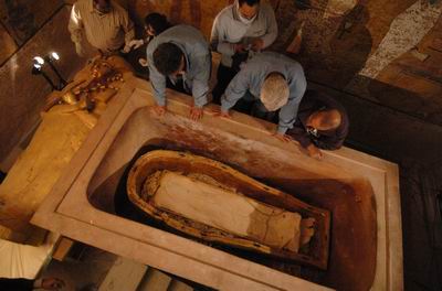
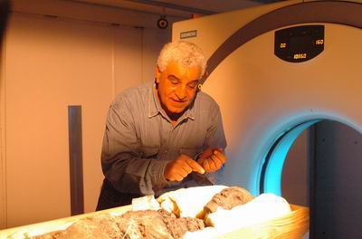
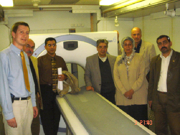
| CT Scan Report On January 5, 2005, the mummy of Tutankhamun (c. 1355-1346 B.C.) was removed from its tomb in the Valley of the Kings (KV 62) for the first time in almost eighty years. An all-Egyptian team, led by Zahi Hawass, lifted the fragile remains, still resting in the tray of sand in which it had been placed by Carter’s team, from their resting place inside the outermost coffin and sarcophagus of the king, and carried them to a state-of-the-art CT scan machine (housed inside a trailer) donated to the Supreme Council of Antiquities by Siemens, Ltd., and the National Geographic Society. The scan took fifteen minutes and produced over 1,700 images. These images were studied by an Egyptian team, under the auspices of Madiha Khattab, Dean of the Faculty of Medicine, Cairo University, and then by a foreign team composed of experts from Italy and Switzerland.
|
|
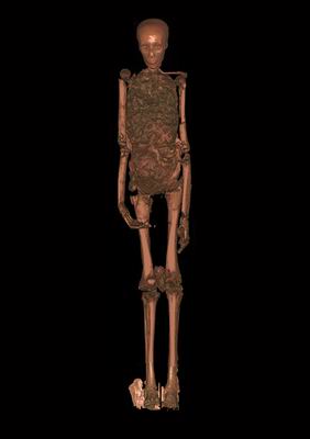
| Previous Examinations The mummy of the young king had been essential dismantled by Carter’s team, who were interested primarily in recovering the almost 150 jewels, amulets, and other items wrapped with the body and gaining the maximum possible scientific information from the body itself. In order to remove the objects from the body and the body from the coffin, to which it was stuck fast by the hardened embalming liquids (most likely resins) used to anoint the mummy, Carter’s team cut the body into a number of large and small pieces (for example, the trunk was cut in half, the arms and legs were detached). The head, cemented by the solidified resins to the golden mask, was severed, and removed from the mask with hot knives. Carter placed the mummy back in the tomb in 1926. The mummy has been X-rayed twice since this time, once in 1968 by a team from the University of Liverpool under R.G. Harrison, and once in 1978 by J.E. Harris of the University of Michigan. |
|
| The Benefits of CT Scanning Although both the original examination and the subsequent X-rays revealed much about the life of the king, they also left many questions open, and have provided fuel for much speculation. CT scanning is a non-invasive tool that can scan the whole body in a very short time, and can differentiate between various types of soft tissue and bone in three-dimensional images. Conventional X-rays can see two planes only and cannot clearly distinguish the soft tissues. The scope and ability of CT scanning to diagnose and differentiate between diseases is also far superior. The body also does not need to be moved repeatedly, as is the case for X-rays. The current investigation was designed to confirm or refute the conclusions of the previous examinations, and look for additional details that earlier investigators might have missed. In this it was extremely successful. |
|
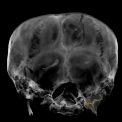
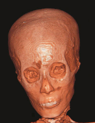
| Results of the CT Scan The scientists who have been working to analyze the CT scan images of Tutankhamun came together in a series of meetings on March 3 and 4 to discuss their findings. The scientists were unanimous on almost all points. Their conclusions were as follows: • State of Mummy. The remains of the pharaoh are in very poor shape, due primarily to the damage done by Carter’s team. The body is in a number of pieces, with both upper and lower limbs dismantled. Many parts present at the original examination are now missing, although many fragments remain loose in the sand tray. Both bones and skin are broken in numerous places. The king’s arms, originally folded across the chest, are now by his sides. • Age at Death. Tutankhamun was about 19 years old when he died, based on the following observations, and using modern developmental tables: |
 |
| a. The fusion of the
epiphyseal plates (the parts of the bone that is responsible for growth
until a certain age) matches the development of a young man of 18 or more,
and 20 or less. b. All of the cranial sutures are still at least partly open. c. The wisdom teeth are not completely erupted. One of these (upper left) is impacted, and there is a slight thinning of the sinus cavity above. This was not life-threatening, and there are no signs of infection. • General Health. Judging from his bones, the king was generally in good health. (His internal organs, as is usual for Egyptian mummies, are not present in the body, and thus have not been analyzed). There are no signs of malnutrition or infectious disease during childhood. His teeth are in excellent condition, and he appears to have been well fed and cared for. • Size in Life. Tutankhamun was approximately 170 cm. (5 and a half feet) tall, as extrapolated from the measurement of the tibia (lower leg). He was slightly built (gracile). • Skull Shape. Tutankhamun had a very elongated (dolichocephalic) skull. The cranial sutures are not prematurely fused, so this is most likely due to normal anthropological variation rather than any pathology. • Cleft Palette and Overbite. The king had a small cleft in his hard palette (the bony roof of his mouth), not associated with an external expression such as a hare-lip or other facial deformation. His lower teeth are slightly misaligned. He has large front incisors and the overbite characteristic of other kings of from his family (the Tuthmosid line). • Scoliosis. There is a slight bend in the spine, However, the scientists agree that this is not a pathological scoliosis, since there is no rotation and no associated deformation of the vertebrae. This bend most likely reflects the way the mummy was positioned by the embalmers. • Brain Extraction. The nasal septa were destroyed by the embalmers, and the brain was extracted through the nose. • Embalming of Head: Principal Route. All the scientist agree, based on the differing densities of the materials and the way in which the embalming liquids (now completely solidified) appear, that various types of these liquids were introduced to the cranial cavity several times through the nose. At first, the body was lying on its back, and the embalming liquid pooled along the back of the skull. Later, the head was tipped back in some way, and embalming liquid pooled in the top of the skull. • Possible Second Route for Embalming of Head. Part the team sees evidence for a second route through which embalming liquid was introduced to the lower cranial cavity and neck. This would have been through the back of the upper neck. In this area, there are two layers of solidified material of a different density from that seen above in this area. The first cervical (topmost) vertebra and the foramen magnum (large opening at the base of the skull) are fractured here, which according to this theory may have happened when the hole was made to pour in the embalming liquid or may have been done by Carter’s team when removing the head from the mask. Part of the team disagrees, and sees no evidence for an embalming route through the back of the neck. They believe, instead, that the embalming liquid in this area was also introduced through the nose or trickled down from the cranial cavity, and that the vertebra and foramen magnum were definitely damaged by Carter’s team in the process of removing the head from mask, and could not have been damaged by the embalmers. • The “Murder” Theory. The entire team agrees that there is NO evidence for murder present in the skull of Tutankhamun. There is NO area on the back of the skull that indicates a partially healed blow. There are two bone fragments loose in the skull. These cannot possibly have been from an injury from before death, as they would have become stuck in the embalming material. The scientific team has matched these pieces to the fractured cervical vertebra and foramen magnum, and believes these were broken either during the embalming process or by Carter’s team. 2. Fractured Leg? The team has noted a fracture of the left lower femur (thighbone), at the level of the epiphyseal plate. This fracture appears different from the many breaks caused by Carter’s team: it has ragged rather than sharp edges, and there are two layers of embalming material present inside. Part of the team believes that the embalming material indicates that this can only have occurred during life or during the embalming process, and cannot have been caused by Carter’s team. They note that this type of fracture, unlike most of the others, is possible in young men in their late teens, and argue that it is most likely that this happened during life. There is no obvious evidence for healing (although there may be some present, and masked by the embalming material). Since the associated skin wound would still have been open, this fracture would have had to occur a short time, days at the most, before death. Carter’s team had noted that the patella (kneecap) on this leg was loose (now it is completely separated, and has in fact, been wrapped with the left hand), possibly suggesting further damage to this area of the body. The part of the team that subscribes to this theory also notes a fracture of the right patella and right lower leg. Based on this evidence, they suggest the king may have suffered an accident in which he broke his leg badly, leaving an open wound. Although the break itself would not have been life-threatening, infection might have set in. However, this part of the team believes it also possible, although less likely, that this fracture was caused by the embalmers. • No Fractured Leg. Part of the team believes that the above scenario is absolutely not possible. They maintain that the fracture mentioned above can only have been done by Carter’s team during extraction of the body from the coffin. They argue that if such a fracture had been suffered in life, there would have been evidence for hemorrhage or hematoma present in the CT scan. They believe the embalming liquid was pushed into the fracture by Carter’s team. • Missing Ribs and Sternum. The sternum and a large percentage of the front ribs are now missing, evidently along with the much of the front chest wall. The ends of the missing ribs are cleanly cut, clearly by a sharp instrument. The scientific team agrees that this cannot mirror in any way extensive trauma to the chest, as such trauma would have been reflected elsewhere in the body (particularly in the vertebra). Opinion among team members is divided as to whether the ribs and sternum were removed by the embalmers or by Carter’s team. Carter’s team does not mention that the ribs and sternum were missing, and a beaded collar and string of beads can be seen covering the chest cavity in photos taken at the time, but before their examination of the body was completed. Therefore it is perhaps more likely that this area of the body, which is now completely missing, was removed by Carter’s team in order to collect the artifacts present (although he does not mention doing so). Archaeological investigation will continue in an effort to resolve this issue. • Embalming Process. The team has concluded, based on the identification of at least five different types of embalming material and the many episodes of its introduction to the body and cranial cavity, that great care was taken in the mummification of this king. This counters previous arguments that the body of the king was prepared hurriedly and carelessly. • Missing Penis. Although they cannot be certain, the team believes that they have located the king’s penis, present at Carter’s exhumation but reported missing at the 1968 examination, loose in the sand around the king’s body. There are also many fragments apparently belonging to other missing parts, such as a thumb, other digits, and pieces of vertebrae, present in this area. |
|
| CT Scanning Team • Egyptologist and Team Leader: Dr. Zahi Hawass, Secretary General, SCA • Project Administrator: Hisham el-Leithy
|
|
| Scientific Team (in
alphabetical order): • Dr. Aly Gamal Eldin, Professor of Forensic Medicine, Cairo University • Dr. Sherief Abdel Fatah, Lecturer in Radiology, Cairo University • Dr. Fawzi Gaballa, Professor of Anatomy, Cairo University • Dr. Ashraf Selim, Professor of Radiology, Cairo University • Dr. Mervat Shafik, Professor of Radiology, Cairo University • Dr. Essam el-Sheikh, Professor of Radiology, Cairo University |
|
|
|
|
| Machine Operators • CT Application Specialist, Seimens, Ltd., Egypt: Dr.Hani Abdel Rahman • Technician: Salah Mohamed Ali |
|
| Scientific Consultants (in
alphabetical order): • Dr. Edward Egarter, Forensic Pathologist, Archaeological Museum of South Tyrol, Italy • Dr. Paul Gostner, Radiologist, General Hospital of Bolzano, Italy • Dr. Frank J. Rühli,Ph.D., M.D. Anatomist, Paleopathologist, University of Zurich |
|