|
PRESS RELEASE, May 10, 2005 |
|
|
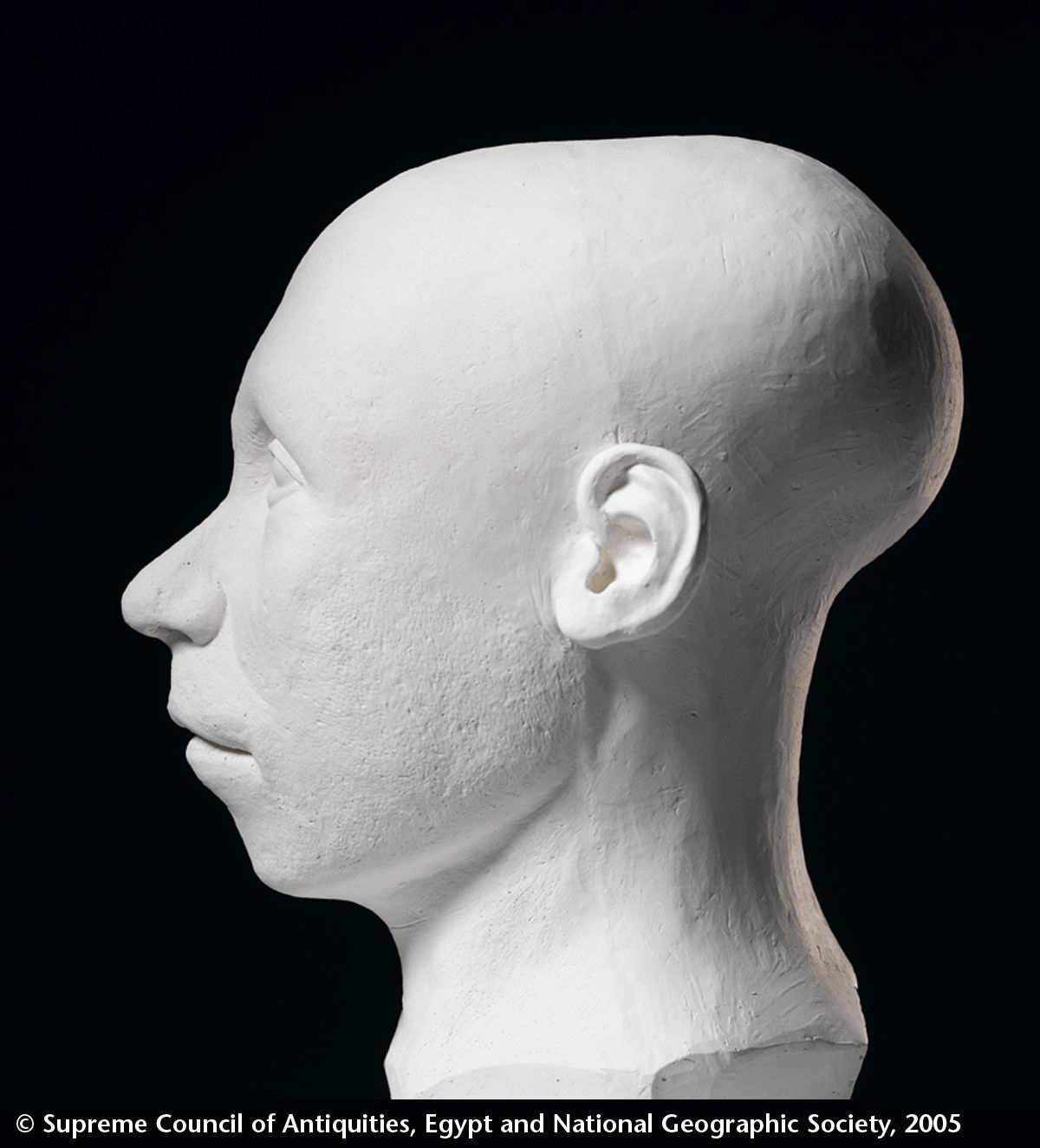
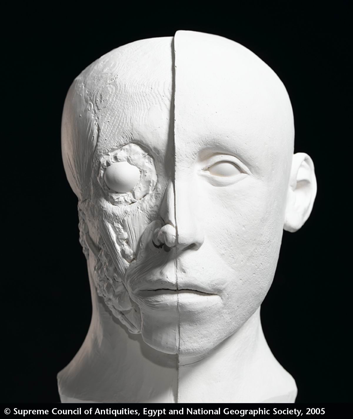
Tutankhamun Facial Reconstruction Dr. Zahi Hawass, Secretary General of Egypt’s Supreme Council of Antiquities, announced today the results of three independent attempts to reconstruct the face of Egypt’s most famous king, Tutankhamun. |
|
|
|
|
|
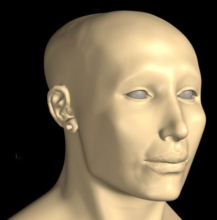
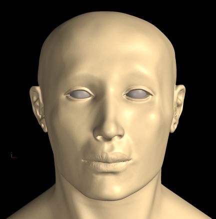
Reconstruction by the Egyptian Team |
|
|
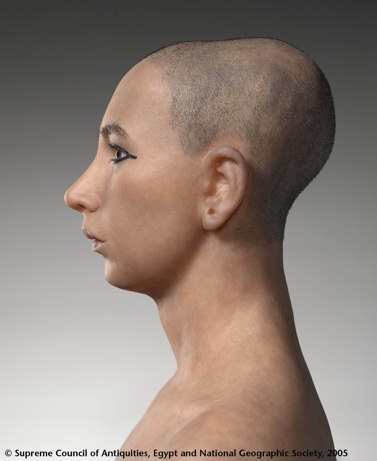
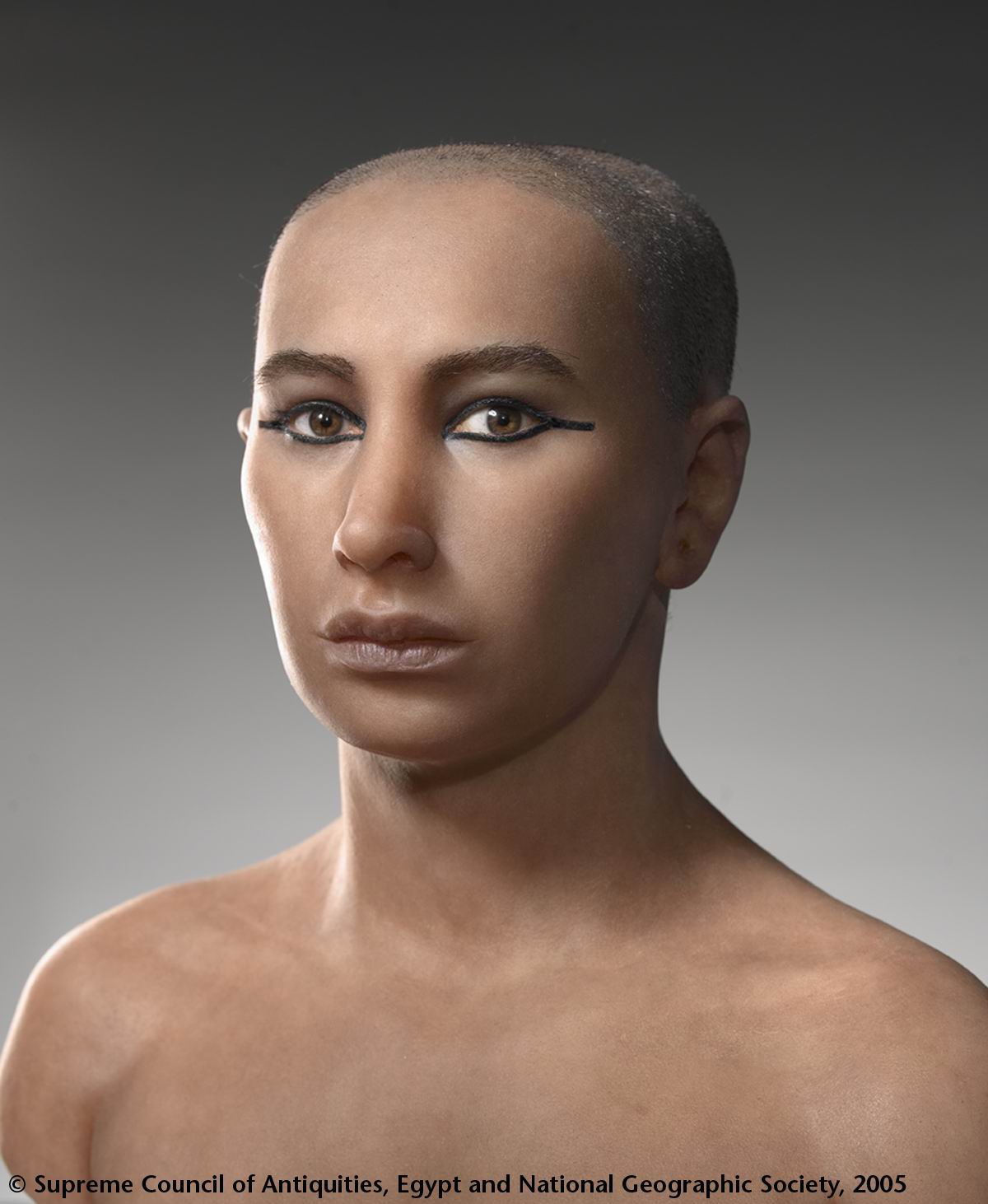
Dr. Hawass led the effort to see what King Tut, who died over three thousand years ago, might have looked like in life. Under his direction three independent artist-scientist teams, one French, one American, and one Egyptian, used modern forensic techniques to reconstruct Tut’s face. Two of these teams were chosen and sponsored by the National Geographic; the third was selected by the SCA. The French and Egyptian teams were told that the subject was Tutankhamun. The American team was not given the subject’s identity and was thus working “blind.” |
|
|
|
|
|
Reconstruction by the Egyptian Team |
|
|
These likenesses are based on CT scan data collected by an all-Egyptian team, led by Dr. Hawass, using a portable CT scanner provided by the National Geographic Society and Siemens AG. The scan took place on January 5, 2005, in the Valley of the Kings in Luxor. The fragile body of the king, which had lain undisturbed since it was last X-rayed in 1978, was carefully carried to the scanner inside the wooden tray of sand in which Carter had left it in 1926. The CT scan was able, with minimum disturbance of the mummy, to distinguish different densities of soft tissue and bone. During the scan over 1,700 high-resolution images were captured; these were then used to create three-dimensional models, both virtual and actual. Results from the analysis of the CT scan were announced on March 5 of this year. The scientific team concluded that Tutankhamun was about 19 when he died. He was well-fed, and showed no signs of childhood diseases or malnutrition. The team found no signs of a blow to the head, as had been theorized based on X-rays taken in 1968, and no other evidence for foul play. They did note a bad fracture just above the left knee that may have occurred a day or two before the king died (rather than being caused by the embalmers or Carter’s team). “It is possible that this injury became infected and killed the king,” says team leader Zahi Hawass. |
|
|
|
|
|
Reconstruction by the American Team |
|
|
All three
teams started with the CT data provided by the SCA. The American and
French teams were given a plastic model of the skull produced in Paris;
the Egyptian team made their own model directly from the data. Based on
this skull, the American and French teams both concluded that the subject
was Caucasoid (the type of human typically found, for example, in North
Africa, Europe, and the Middle East). The American team, working blind,
correctly identified the subject as North African. |
|
